A Colombian woman has given birth to a baby whose abdomen contained the tiny, half-formed — but still growing — body of her own twin sister.
This type of birth, an example of “fetus-in-fetu,” is very rare but not unprecedented.
[Like the Science Times page on Facebook. | Sign up for the Science Times newsletter.]
The condition was described in a British medical journal in 1808 and is thought to occur in about one in every 500,000 births. In recent years, similar births have occurred in India, in Indonesia and in Singapore.
The latest case was even more unusual, because doctors clearly identified the fetus-in-fetu during the pregnancy, said Dr. Miguel Parra-Saavedra, a high-risk pregnancy specialist in Baranquilla, Colombia, who oversaw the birth.
He first saw the mother, Monica Vega, when she was in her 35th week of pregnancy, five weeks short of a full-term birth. Her obstetrician believed her fetus had a liver cyst.
But, using color Doppler and 3D/4D ultrasound imaging, Dr. Parra-Saavedra was able to see that the fluid-filled space actually contained a minuscule infant, supported by a separate umbilical cord drawing blood where it connected to the larger twin’s intestine.
“I told the mother, and she said, ‘What? No, doctor, this is impossible,’” Dr. Parra-Saavedra said. “But I explained step by step, and she understood.”
He alerted a local television news network, which followed Mrs. Vega, who is now 33, through the birth of her daughter, Itzamara, and the surgery to remove Itzamara’s partially formed twin.
(Dr. Parra-Saavedra had developed relationships with several journalists during Colombia’s Zika outbreak in 2016, because he treated mothers whose babies had microcephaly, or small heads, caused by the virus.)

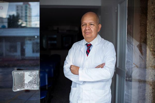
Dr. Miguel Parra-Saavedra, a high-risk pregnancy specialist in Barranquilla, Colombia, who delivered Itzamara at 37 weeks.CreditKatie Orlinsky for The New York Times
On Feb. 22, when Itzamara was at 37 weeks and weighed about seven pounds, doctors decided to deliver her by cesarean section, because they feared the internal twin would crush her abdominal organs.
The next day, they removed the fetal twin by laparoscopic surgery. It was about two inches long and had a rudimentary head and limbs, but lacked a brain and heart, Dr. Parra-Saavedra said.
Fetus-in-fetu is sometimes misdiagnosed as a teratoma, a tumor that may contain bones, muscle tissue and hair. A DNA comparison is being done, but Dr. Parra-Saavedra has no doubt that the fetuses started out as identical twins from the same ovum.
Because the smaller fetus took nourishment from its sibling, it is called a heteropagus or parasitic twin.
Some heteropagus twins are born conjoined to their healthy siblings, while some grow partially inside and partially outside their twin’s body.
The fetus-in-fetu condition is believed to arise soon after the 17th day of gestation, when the embryo flattens out like a disc and then folds in on itself to form the elongated fetus.
Doctors believe that in exceedingly rare cases, the twin embryos only partially divide, and the larger one wraps around the smaller.
The condition may go undetected for many years. In 2015, a 45-year-old Englishwoman living in Cyprus underwent surgery for what appeared to be a four-inch tumor on her ovary.
On dissection, the growth turned out to have a partially formed face with an eye, a tooth and black hair. Doctors concluded that it was a twin she had absorbed while in her mother’s womb.
Itzamara is doing well, Dr. Parra-Saavedra said. “She has a little scar on her abdomen, but she is a normal baby now except that the whole world is talking about her.”

